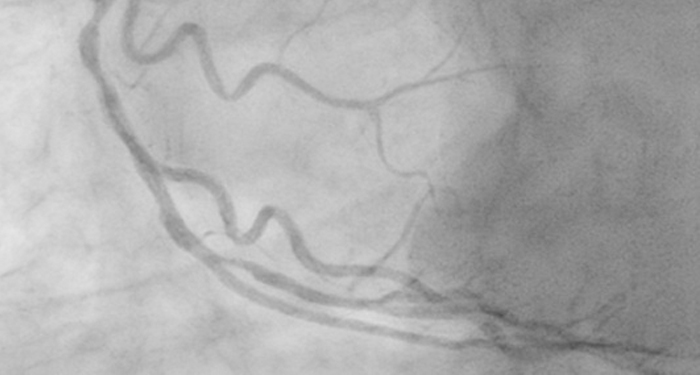
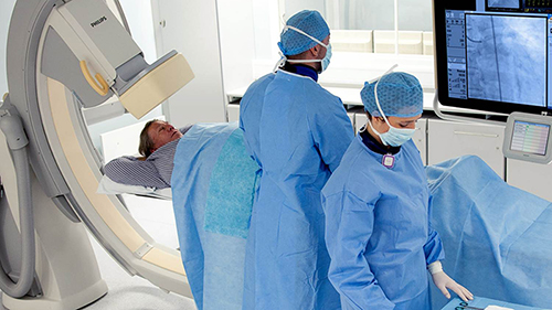
Live Image Guidance in treating structural heart disease
New, complex SHD procedures require specialists to work together, with guidance from X-ray and echo for visualization of critical anatomy. Whether it’s TAVR, Mitral valve clipping, or LAA occlusion, 3D imaging enhances identification of target points, simplifies device deployment, and offers immediate evaluation of results. Philips Live Image Guidance helps optimize real-time navigation through soft tissue anatomy. Procedure specific tools and integration of imaging modalities boosts confidence and enhances workflow among the heart team. Sign up to receive exclusive updates on Live Image Guidance.
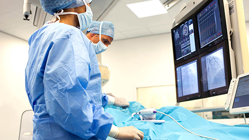

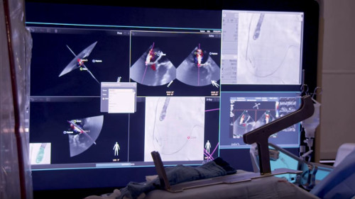
Greater insight and confidence in finding and treating the problem
Our Live Image Guidance expands clinical capabilities through intelligent and intuitive integration of multi-modality images at the point of treatment, enabling confident diagnoses and real-time therapy monitoring.
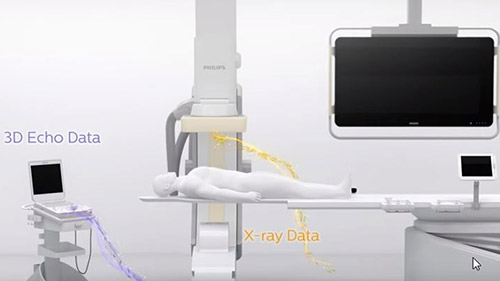
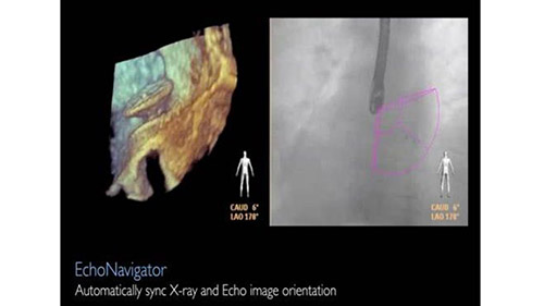
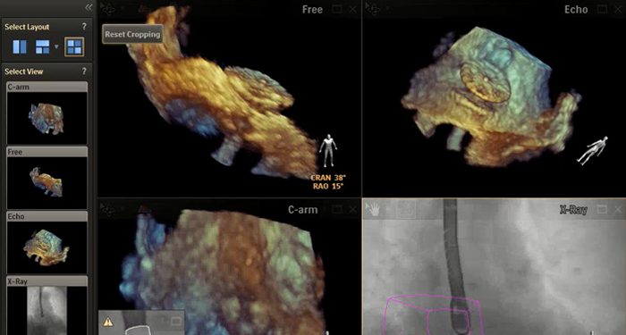
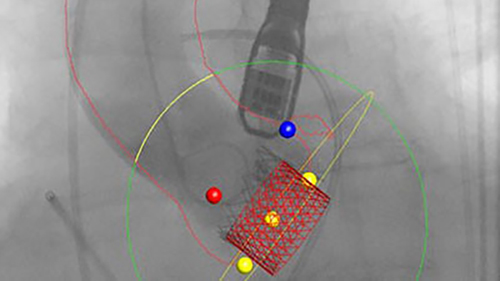
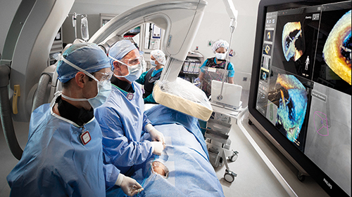
EchoNavigator
Real-time, automatic, fusion of live X-ray & live echo
By automatically fusing live X-ray and live echo images, in real time, EchoNavigator helps you identify target points and provides enhanced navigation of devices through soft tissue anatomy during interventions.
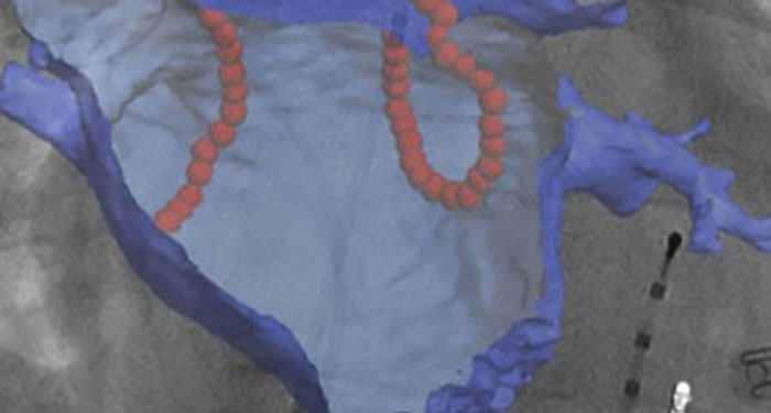
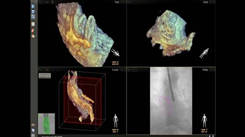
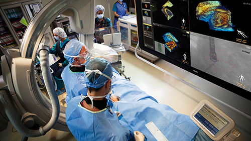
HeartNavigator
3D live image guidance
HeartNavigator provides automatic segmentation and landmark identification, for simplified TAVR planning and device/projection selection. Assessment of calcification around the valve helps to avoid procedure complications.
Better user experience to promote consistency and efficiency
By combining disease centric tools, patient data, and procedural protocols in a single suite, clinicians are supported in each of their specific disciplines. This simplifies clinical workflow and promotes efficiency.
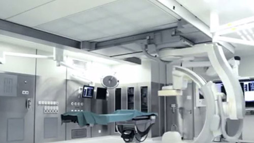
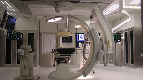
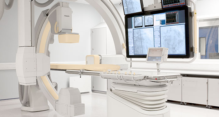
Hybrid Suite with FlexMove option
Versatile room setup for interventionalists
Ceiling mounted X-ray system allows the angio unit to be positioned to either side of the table for easy access to patient. It can be moved aside or completely out of the way when desired.
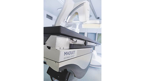
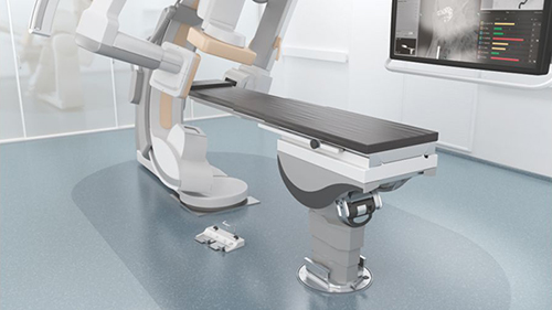
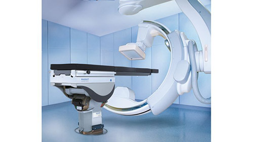
MAQUET table
Improved hybrid suite functionality
Flexible design with exchangeable tabletop allows for interventional treatment of structural heart disease as well as open surgery. The ability to place patients in virtually any position broadens the range of procedure options.
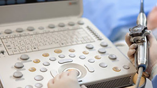
CX50 xMATRIX
Compact ultrasound with 3D TEE
Live 3D TEE and ICE imaging offers increased visibility during guided catheter procedures. The CX50 integrates seamlessly to help speed preparation and reduce duplication through automatic transfer of patient data from X-ray to ultrasound system.
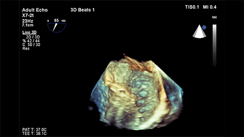
EPIQ
Powerful ultrasound performance
Captures an enormous amount of acoustic data and reconstructs in real-time optimally focused beams, creating precise resolution for every pixel in the image. You get superb tissue uniformity and detail.
FlexVision XL
Full control of your viewing environment
Integrates all relevant images and information together in an interactive, intuitive and procedurally relevant way to expand clinical capabilities and enhance real time cross functional communication.
Lower barriers for minimally invasive interventions
An innovative set of techniques, programs, and practices are used to deliver high quality imaging for a full range of clinical procedures at ultra low dose. This enables comfortable treatment of radiation-sensitive patients and helps manage staff exposure.
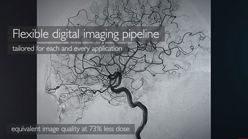
AlluraClarity with ClarityIQ
Industry leading image quality at a fraction of the dose
AlluraClarity family of X-ray systems with ClarityIQ technology delivers superb image quality at 50% dose reduction, helping to minimize patient and staff exposure. You can manage low dose levels to see what you must, without changing the way you work.
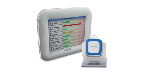
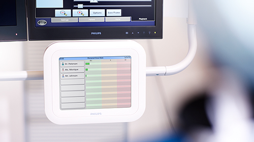
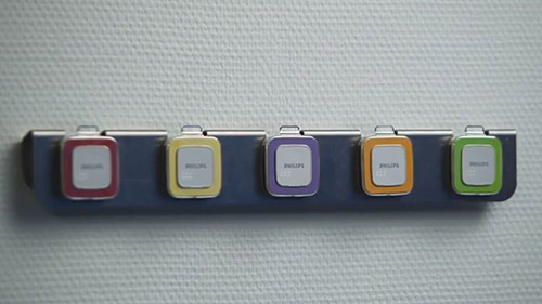
DoseAware family
Real-time radiation exposure monitoring
Real-time dose feedback tools include DoseAware Xtend for procedure-based feedback on scattered X-ray dose (during and after the procedure) to help manage radiation exposure for patients and staff.
Increased economic value
We are committed to working with you to reduce re-admissions, streamline work flow, and increase patient volume by opening the door to new procedures and techniques. By supporting a wide range of procedures you can increase system utilization and reduce the total cost of ownership. Flexible service contracts protect your investment over its entire lifecycle, increasing uptime and providing easy access to upgrades and innovations.